Keep a clear view thanks to our ophthalmoscopy devices
Ophthalmoscopy - also known as funduscopy or eye examination - is a well-known procedure in the eye care medical field. This examination consists of examining the eye’s fundus, or, to be more precise, the eyeball’s inner surface, with an ophthalmoscope. The ophthalmologist looks through the pupil into the eye’s interior, illuminating it with a light source. The retina, the choroid, and the optic disc are clearly visible thanks to the light and can be therefore well examined. There are two different kinds of ophthalmoscopy procedures: the direct one and the indirect one.
Direct ophthalmoscopy
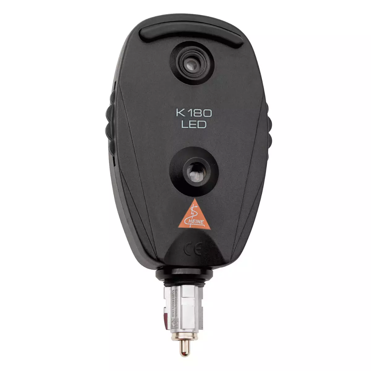
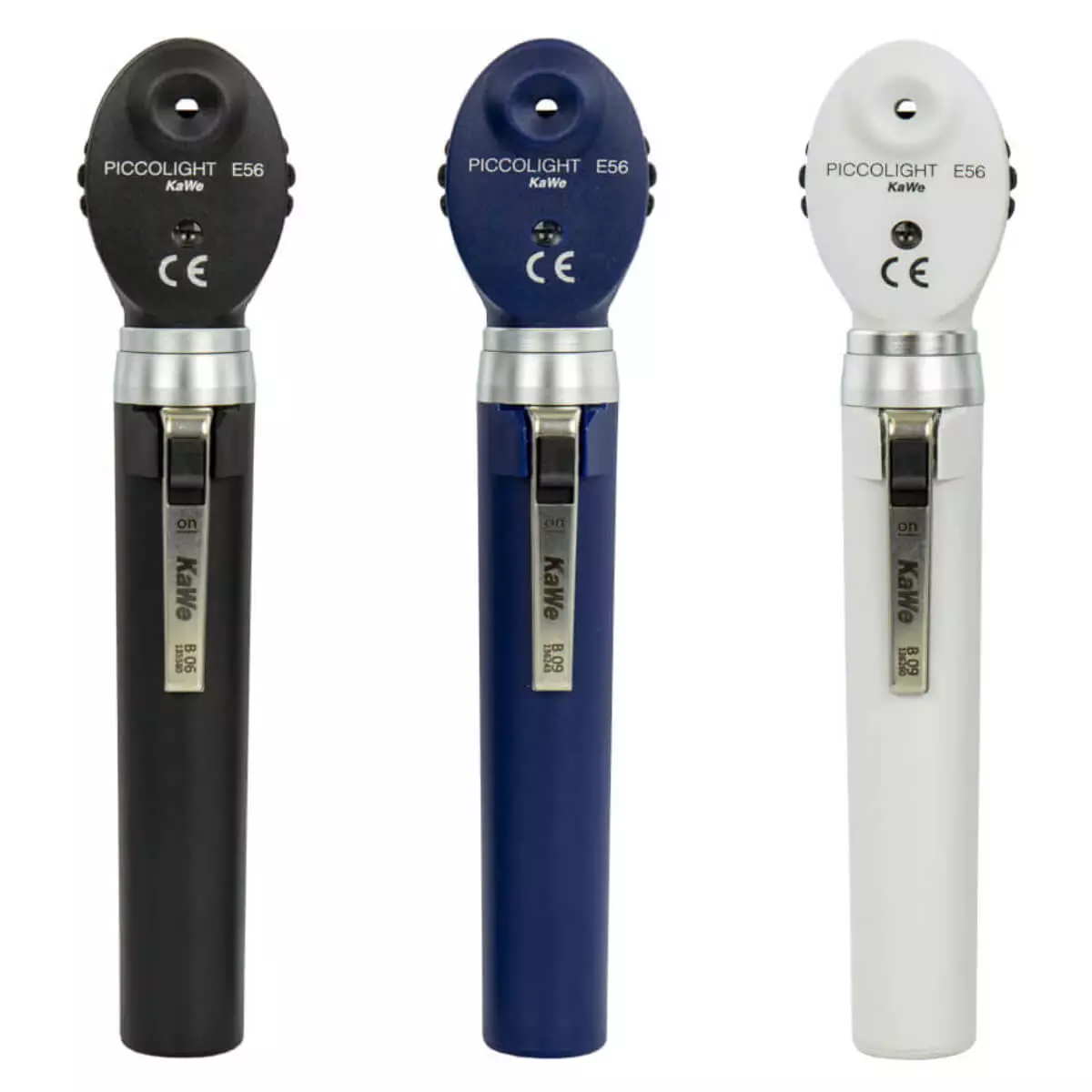
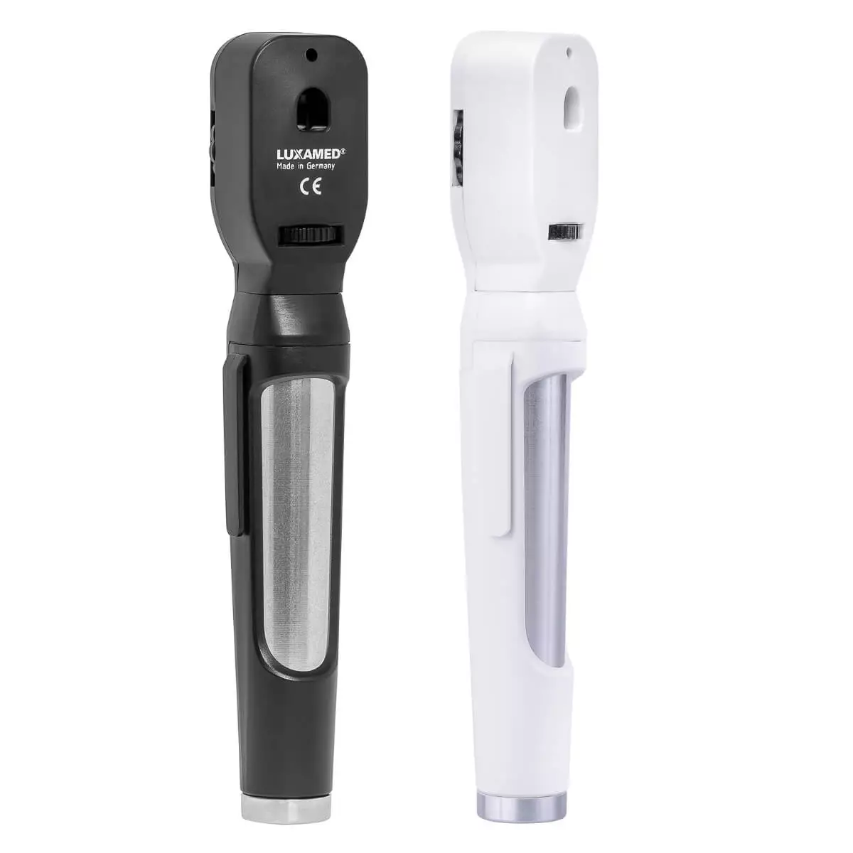
Direct ophthalmoscopy means the doctor uses an electric, hand-held ophthalmoscope. The doctor looks into the patient’s eye with a 14x/16x magnification thanks to the diagnostic instrument. This method is relatively easy to perform and allows you to examine the optic nerve, the macula and the central blood vessels. A disadvantage of direct ophthalmoscopy, however, is that it only shows a small part of the eye’s fundus, although with a high magnification.
Indirect ophthalmoscopy
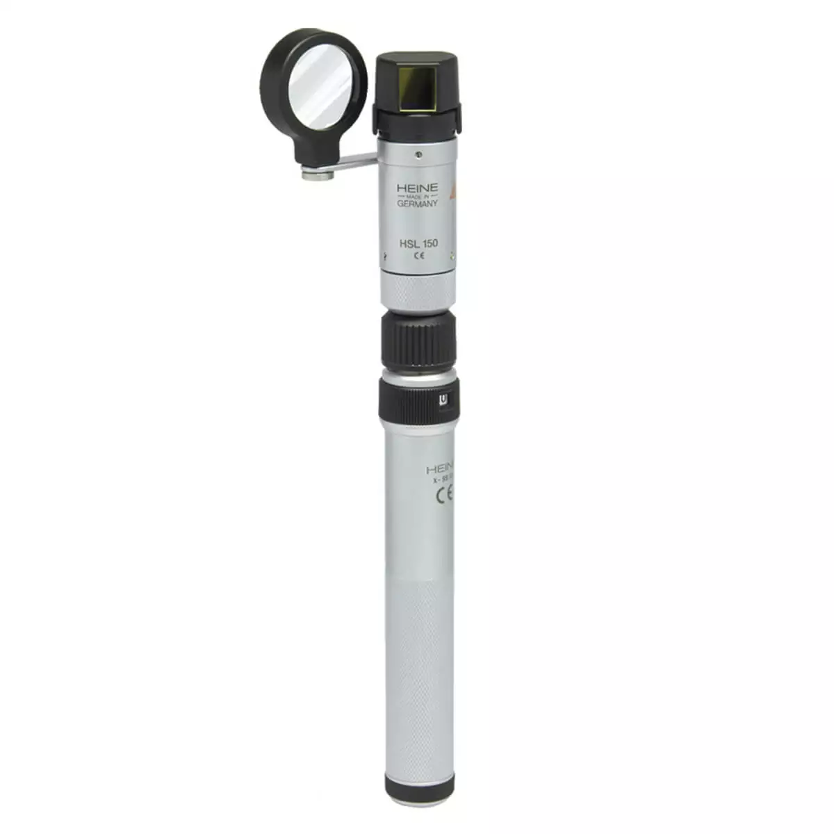
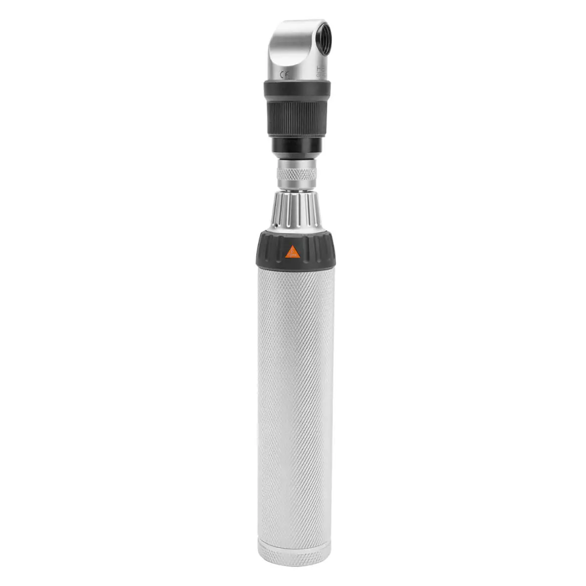
During an indirect ophthalmoscopy procedure, the patient looks above the doctor’s head while they hold a condensing lens in front of them. The doctor rests the arm with the condensing lens on the patient’s head and holds in their other hand a light source which additionally illuminates the eye. Having a good illumination is vital, since it makes the small vessels visible. This method allows you (unlike the direct one) to perfectly see the eye’s entire fundus, however, only with a 4.5x magnification. The image is also upside down, which is why people also call this method an “inverted image” ophthalmoscopy. To make sure you don't run out of light during the examination, you should definitely take a look at our battery & charging handles category. Here, you will find you may need during your next ophthalmoscopy.
Limitations and impediments
It is not possible to use an ophthalmoscope on all patients. Some pathologies like corneal/lens opacity or vitreous hemorrhages prevent you from getting a clear view of the fundus. In cases like these, you can dilate the pupil with some special drops, which will help you see the retina’s peripheral areas better.
Ophthalmoscopes at DocCheck Shop
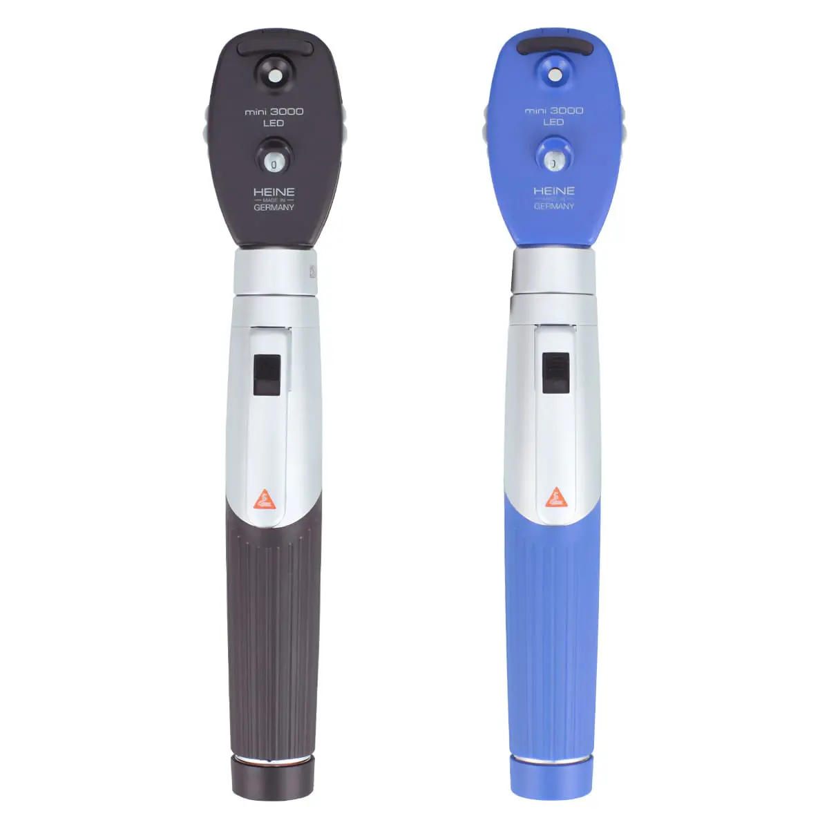
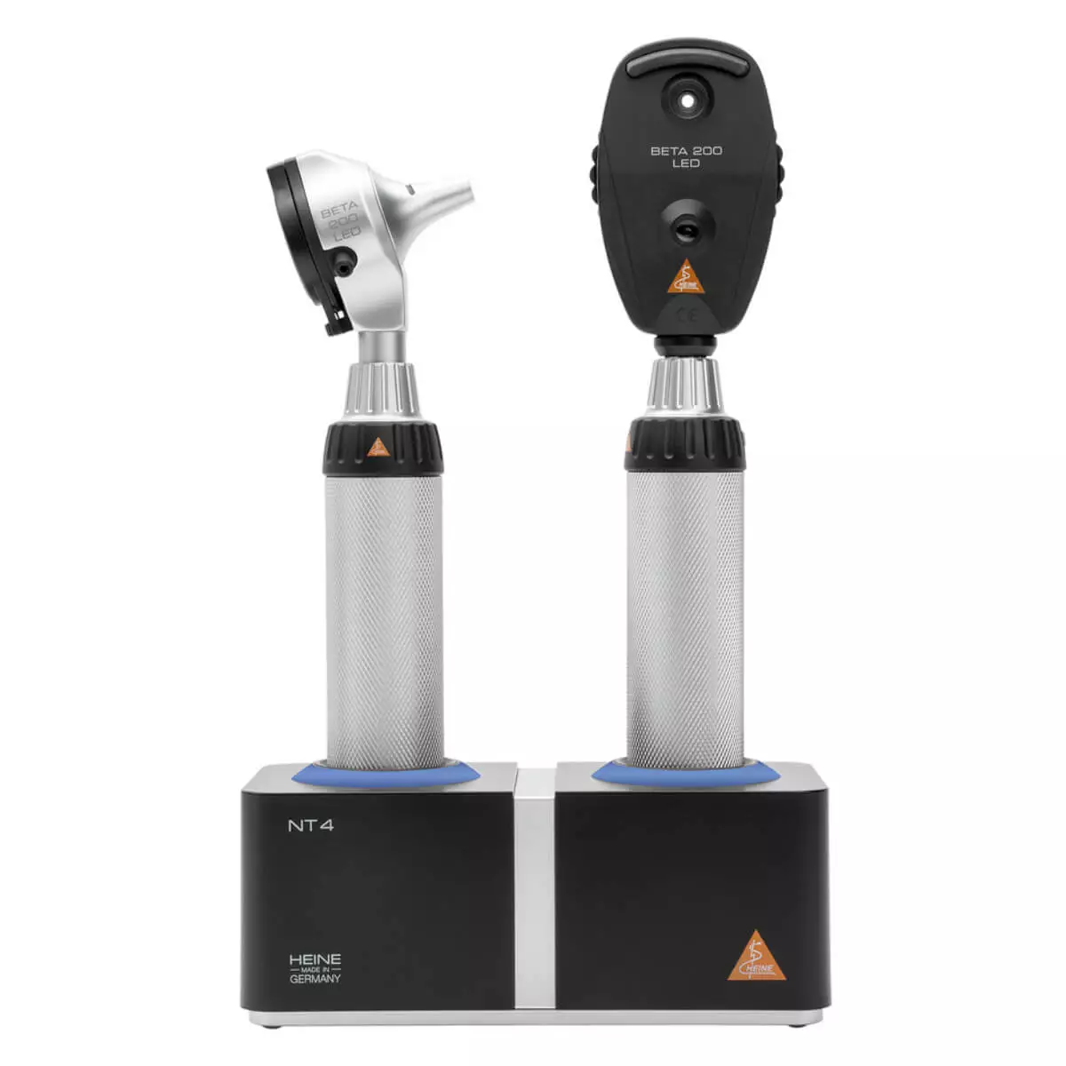
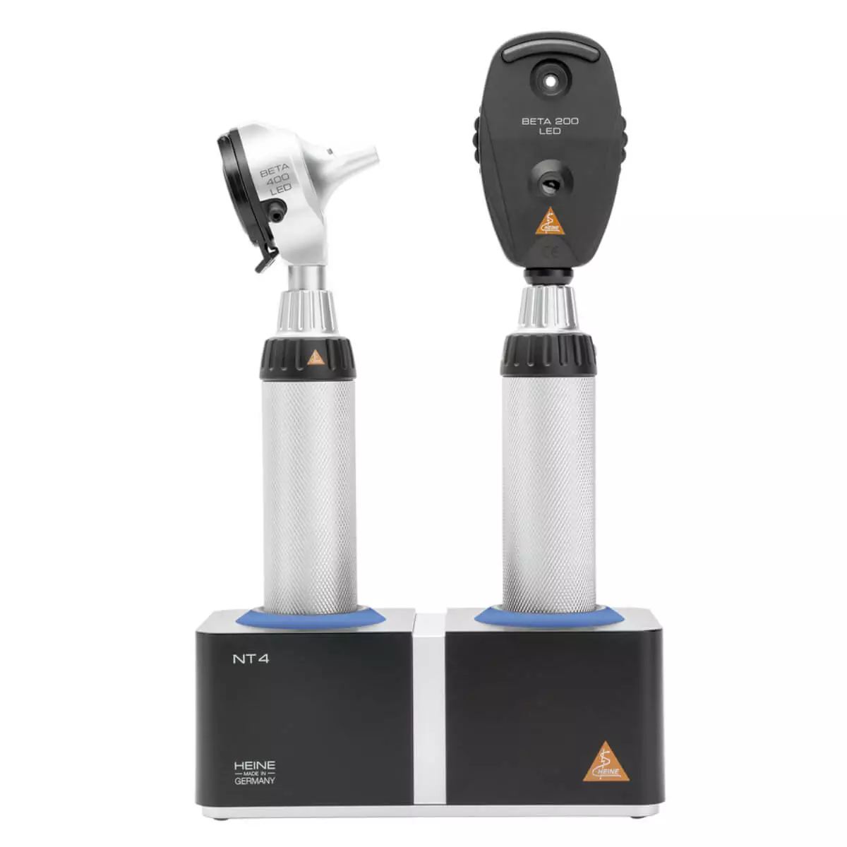
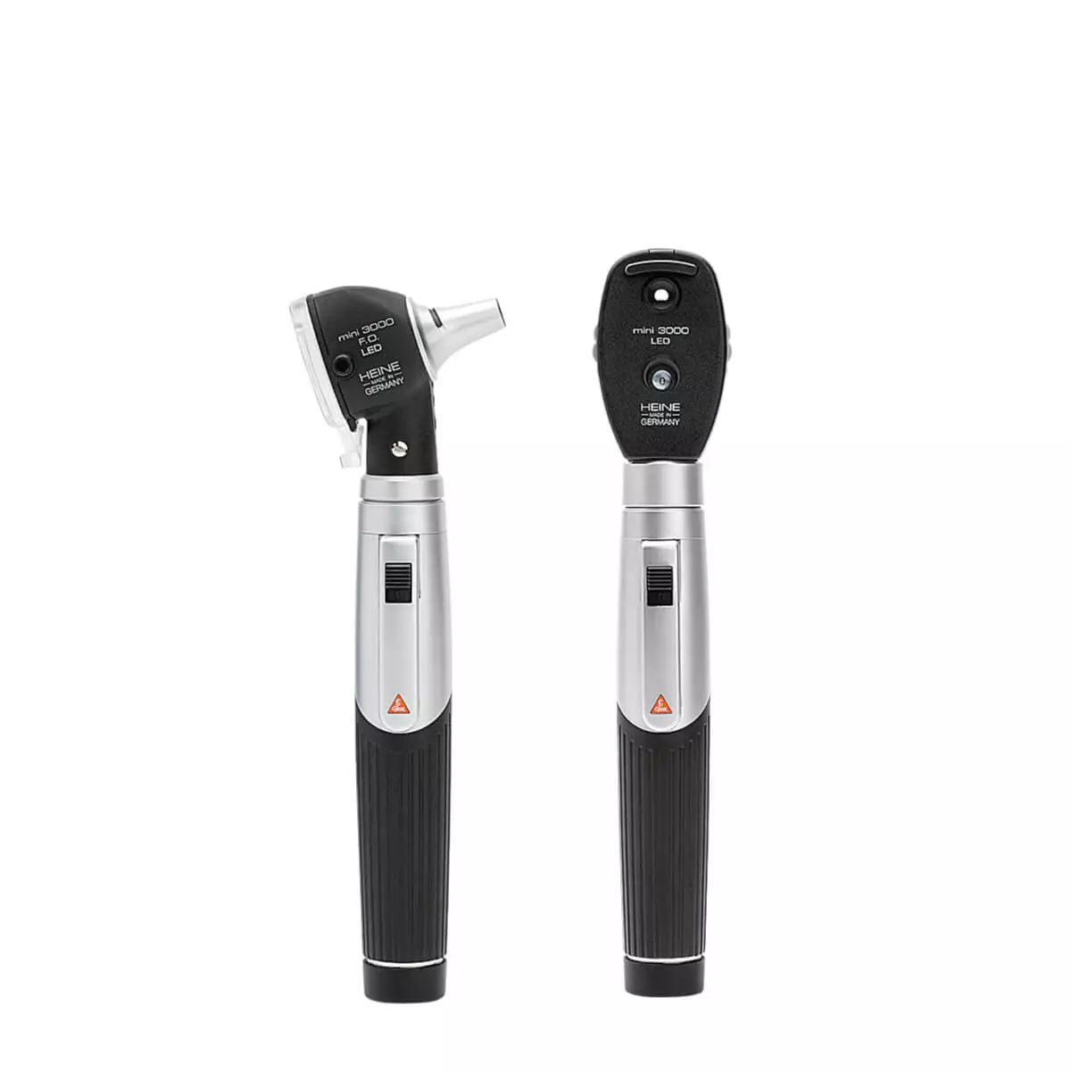
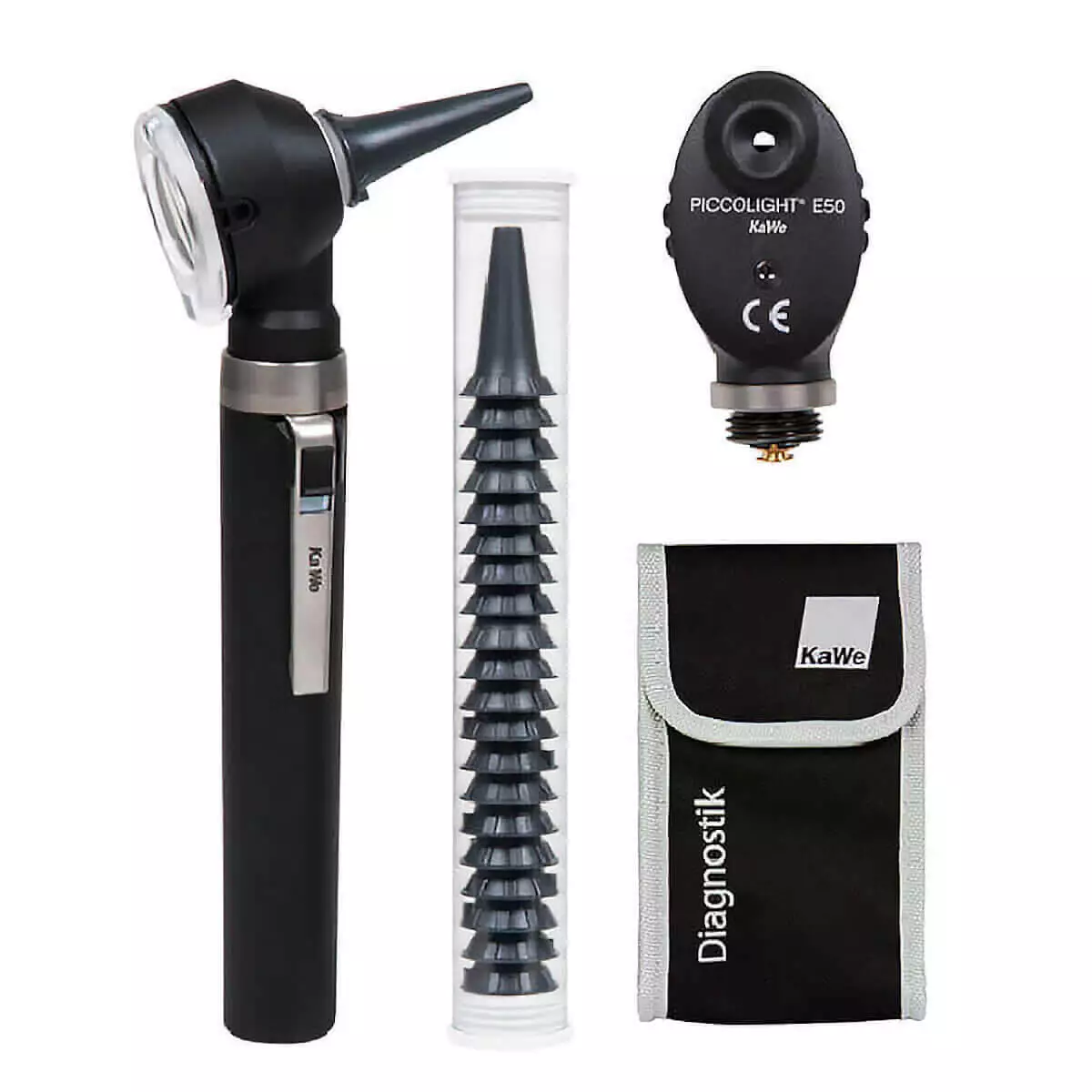
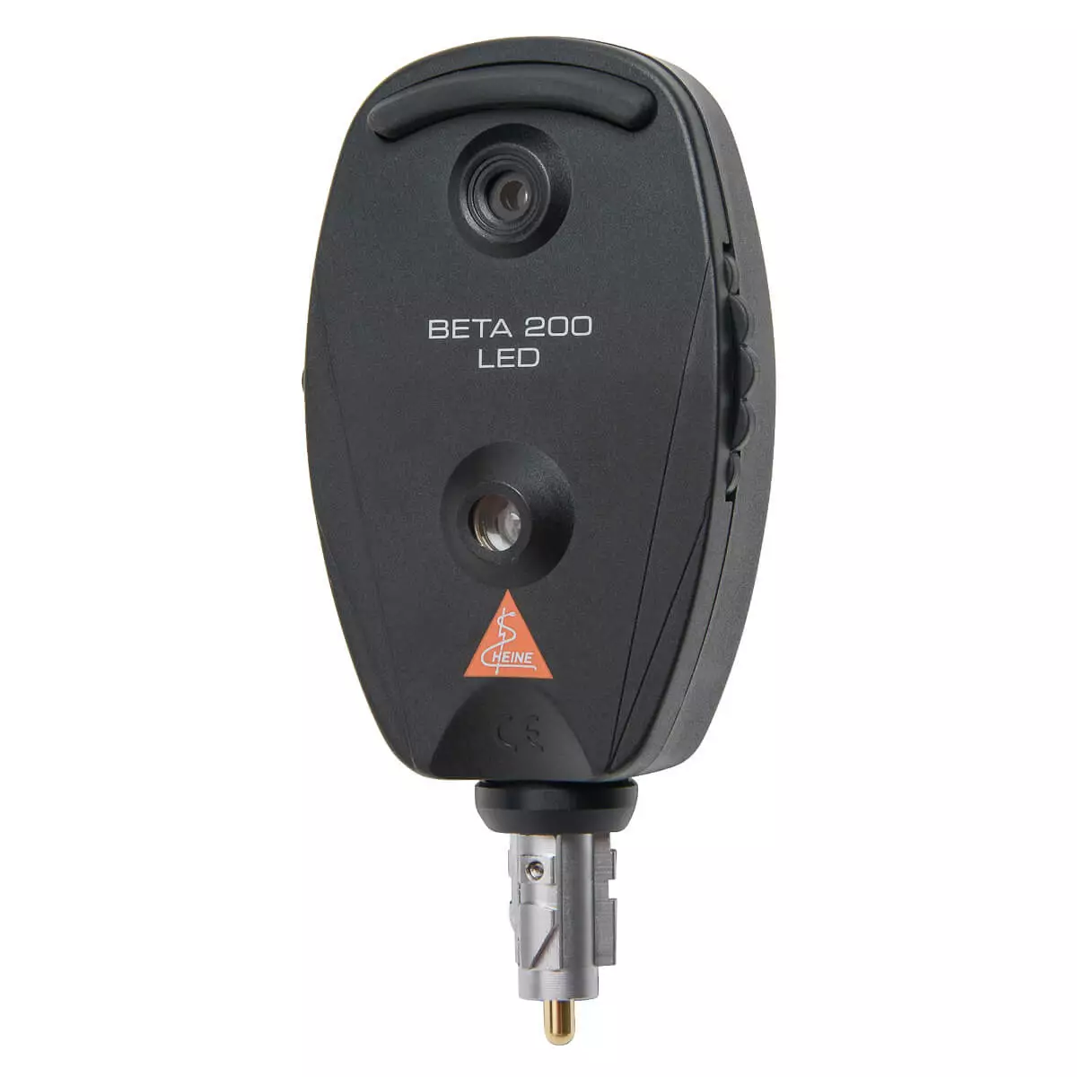
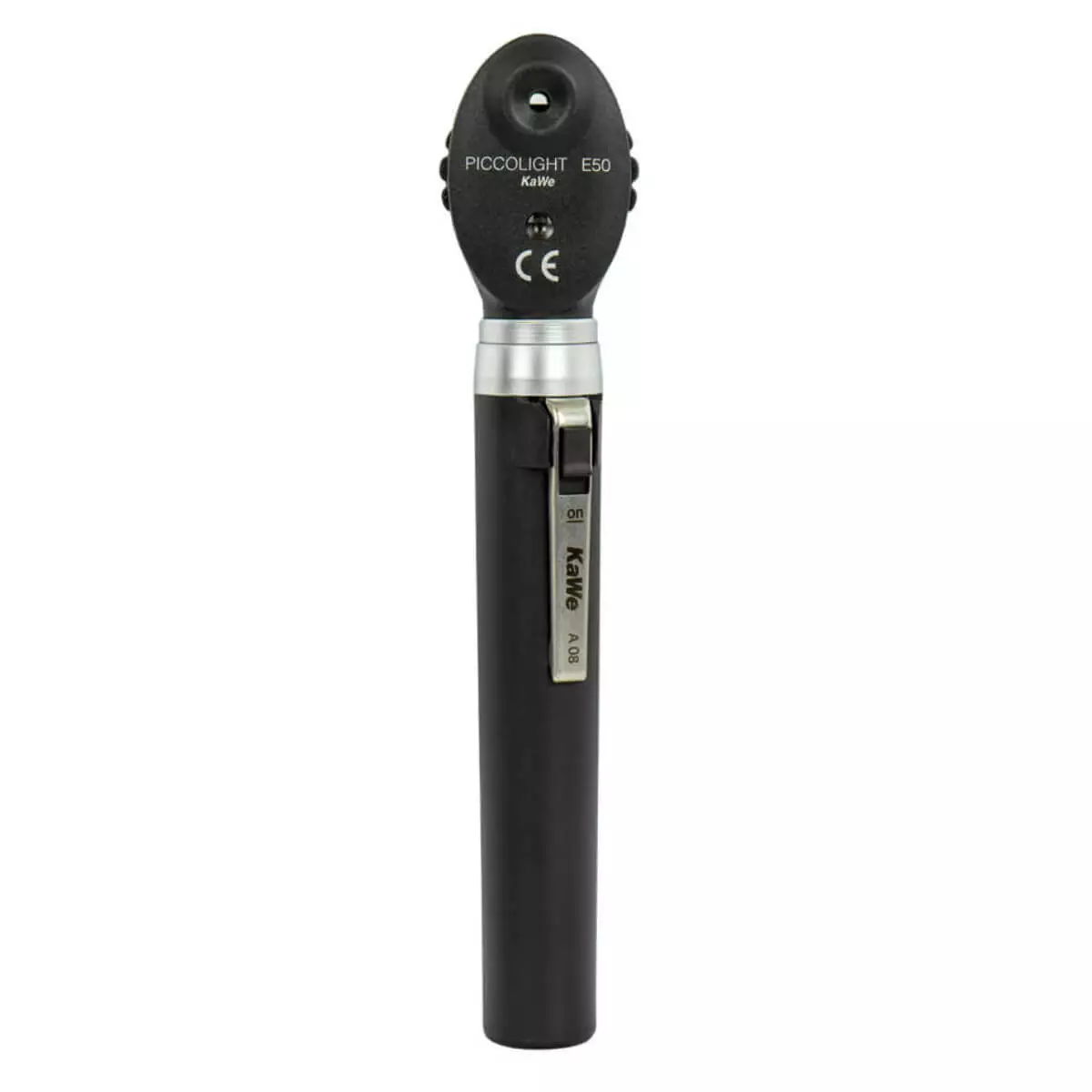
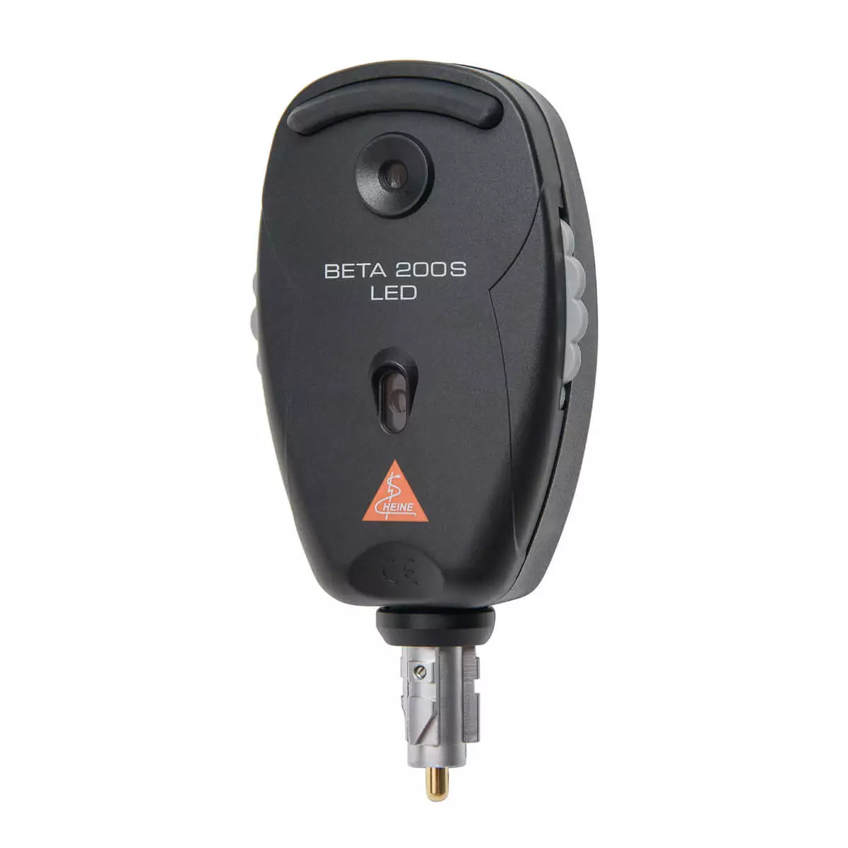
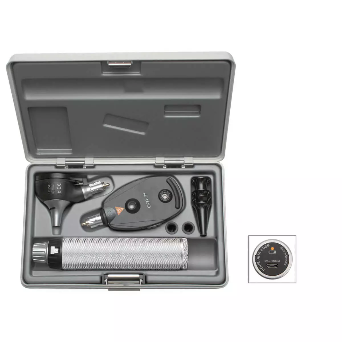
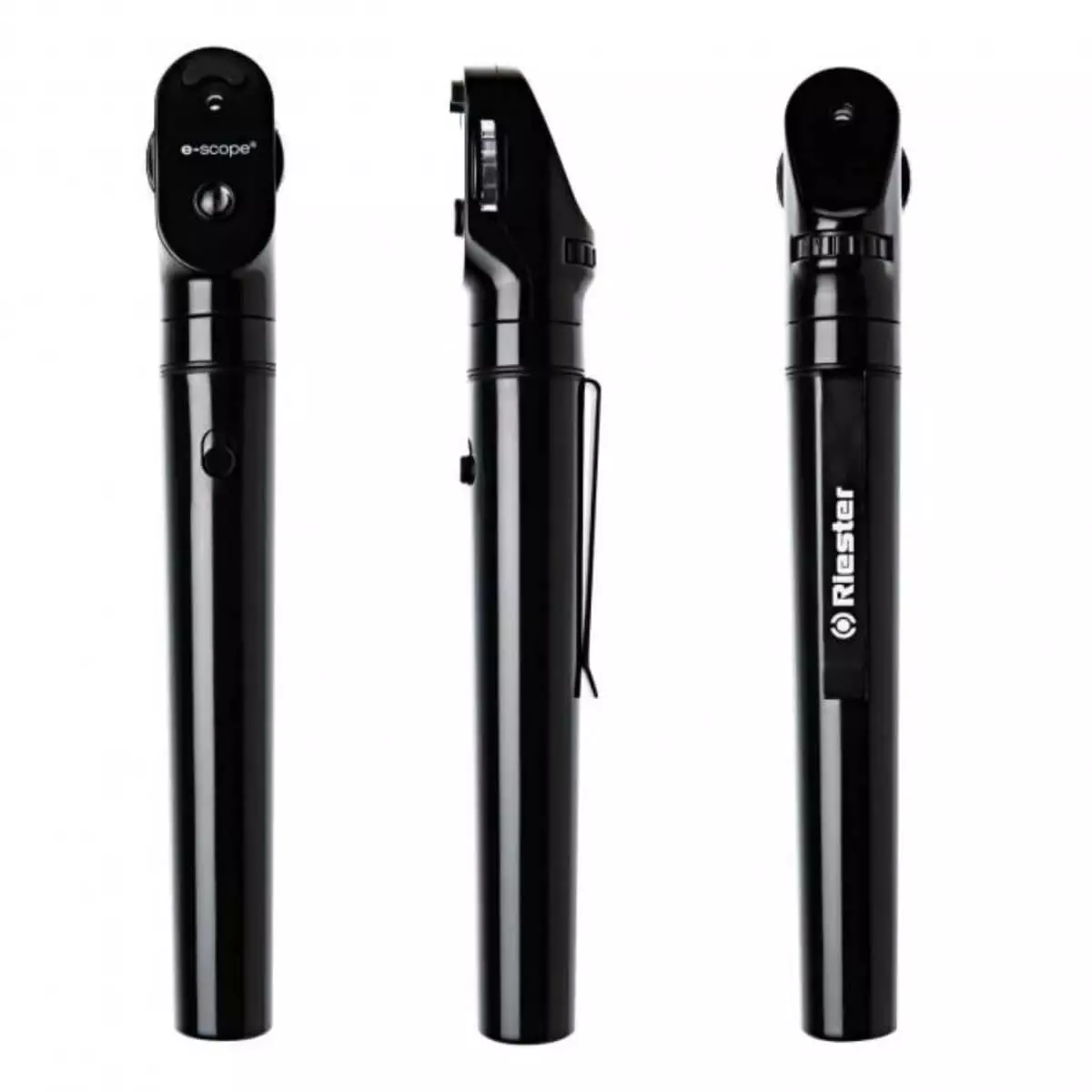
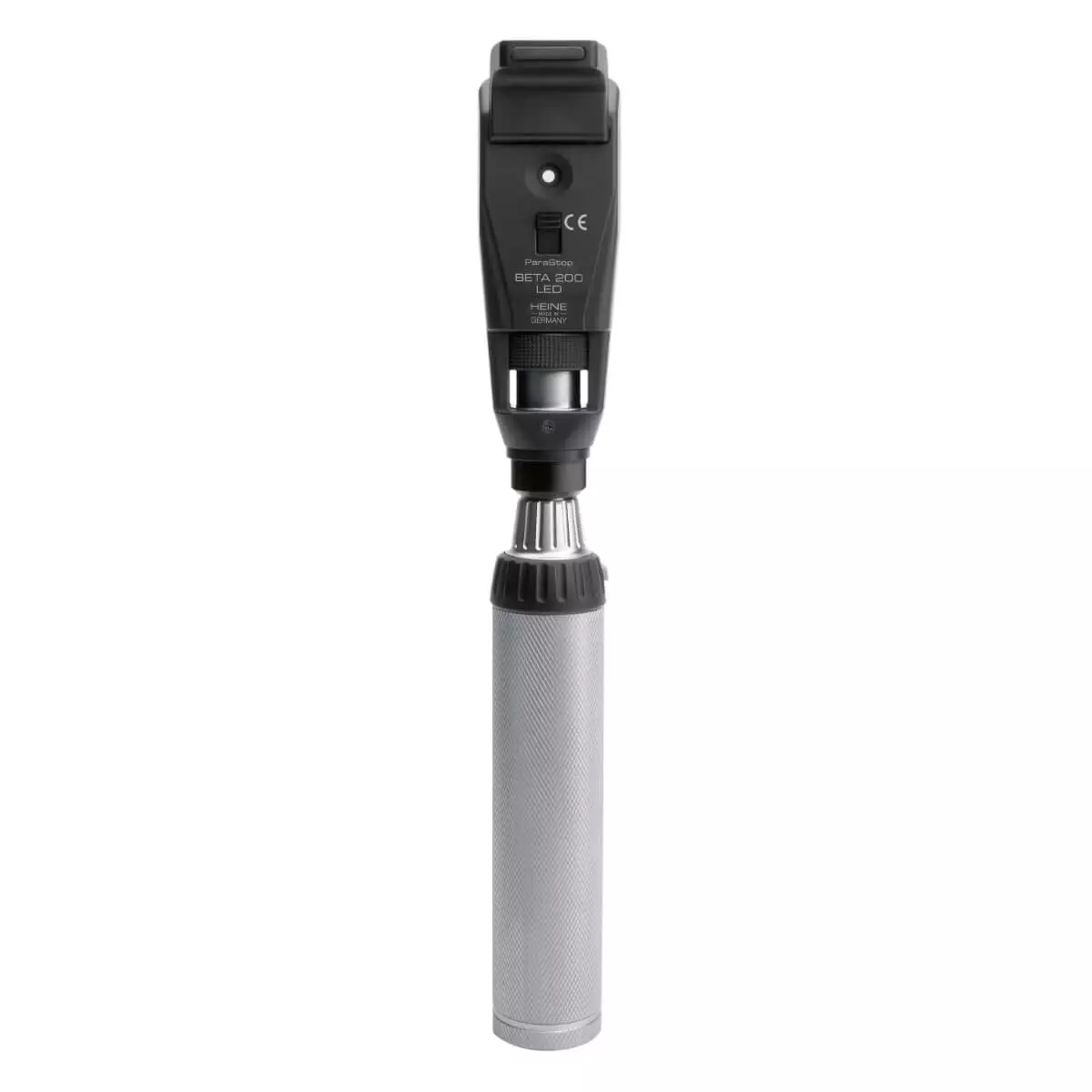
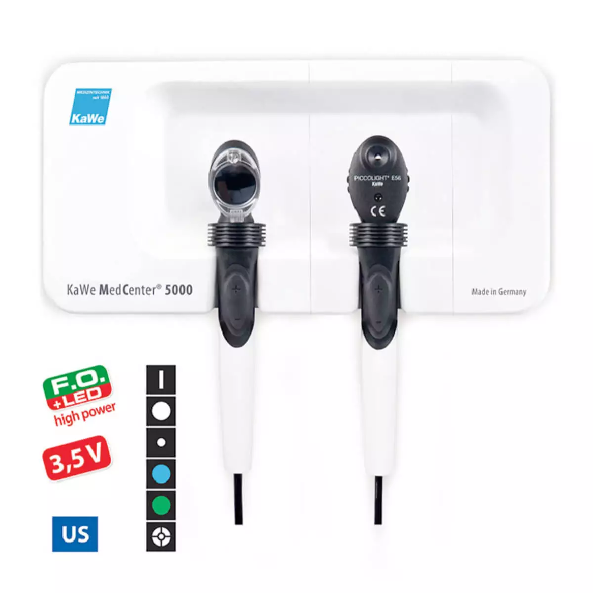
DocCheck Shop is the right place to purchase ophthalmoscope for your next funduscopy. When looking for the right one, you can choose between instruments from brand manufacturers like Heine, for example the BETA 200 LED ophthalmoscope set, which you will find on the diagnostic sets category. We have ophthalmoscopes in different sizes – some for the clinic, some pocket-sized for home visits. We also have devices with different illumination kinds: fibre optic illumination, halogen, energy-saving LED illumination… Go to our ophthalmoscopes category and take a look at our products.


