
140 years of expertise

4,8/5 stars (> 1000 reviews)

Fast delivery
Education


The knee joint model is an anatomical model of a human knee joint with ligamentous apparatus. The full-size knee joint model is firmly mounted on a white plastic base with stand. In addition, the knee joint model is movable, which makes it even easier for the doctor to explain to the patient the processes in the knee joint and the function of the ligaments and tendons during movement. Product details Movable knee joint model Life-size With white plastic base on stand The following structures, among others, are depicted on the knee joint model- Fibula- Tibia- Patella- Femur- Patellar tendon- Inner and outer meniscus Material: PVC Dimensions without base: W 10 x H 30 x D 8 cm Scope of delivery 1 Anatomical knee joint model


The 40-piece torso model shows the anatomy of both sexes in life size. Both the back section with muscle layers, removable vertebrae and nerves, as well as the abdominal side with open neck and all internal organs are represented in detail by the torso model. Since the individual structures are color-coded, the model torso is particularly suitable for learning human anatomy in studies and training. The model torso is not only used for demonstration in an educational context, but also to support patient discussions. A variety of possible diagnoses and forms of treatment, as well as planned interventions, can be vividly explained. Product details 40 parts in exact, detailed representation Life-size, systematically and color-coded Ideal illustrative material in school and university as well as in patient discussions Opened back: Muscle layers, nerve branches and spine with two removable vertebrae to show spinal cord, spinal nerves, 3 meninges Open head: exposed brain Open neck: visible organs and structures Male and female genitals interchangeable Stomach can be disassembled in two parts Mounted on sturdy base Height without base: 85 cm Weight: 10 kg Scope of delivery 1 bisexual model torso

The anatomical eye model consists of 6 separable parts in 3 times life-size. The eyeball can be divided into 2 parts to make the internal structures visible. For example, the lens and vitreous body can be removed from the eye model and viewed more closely. Thanks to the clear, color-coded representation of the ocular membrane, eye muscle attachments, retina with the yellow spot, optic nerve and blood vessels, the model eye is ideally suited for training and study as well as for patient education. Please note: The eye model may differ slightly from the model shown above. Product details 6-part model with many details 3 times life-size Eyeball with muscle attachments and optic nerve Carefully painted structures Vitreous body, lens, iris and cornea can be removed individually Incl. plastic base Dimensions (with base): H 28 x W 12 x D 12 cm In optometry, optics, ophthalmology: eye models Anatomical eye models enable a comprehensive and precise examination of the human eye. Both trainee doctors and opticians benefit from the exact, enlarged and color-coded representation of the human eye. As the model can be broken down into individual parts, it offers a comprehensive insight into the human anatomy. Apart from study and training, the eye model is also an attractive addition to opticians' and doctors' surgeries. Package contents 1 anatomical eye model


In this model, the skull was removably mounted on the flexible cervical spine. The rhomboid brain, spinal cord, cervical nerves, vertebral arteries, basilar artery and posterior cerebral arteries are shown. On tripod. high quality original impression hand-made from hard, unbreakable plastic material very accurate representation of fissures, foramina, processus, sutures and other details can be disassembled into skullcap, skull base and mandible Dimensions: 20 x 13,5 x 15,5 cm Weight: 0,6 kg

In this dental skull, the lower jaw is chiseled open, exposing the tooth roots with vessels and nerves. Skull bones, bone elements, fissures, foramina and other structures are numbered. The cranial sutures are shown in color, as are meningeal vessels and venous blood conduits (sinuses). high quality original impression handmade of hard, unbreakable plastic material very accurate representation of fissures, foramina, processus, sutures and other details can be disassembled into skullcap, skull base and mandible optionally you can insert a 5-part brain (C18) Dimensions: 20 x 13,5 x 15,5 cm Weight: 0,7 kg

So robust, you'll never need another spine again! The special mounting of the 3B Scientific spine model on a flexible metal core ensures particular robustness - making the model especially suitable for the heavy use of regular and active teaching purposes. For the most descriptive demonstrations, the anatomy model is flexibly movable and also features a dorsolateral disc herniation. Product details Detailed teaching model with male pelvis Ideal for regular teaching purposes and heavy use Also suitable for patient education Flexible design for illustrative demonstrations Particularly robust due to stable mounting on flexible metal core With occipital scales, vertebral arteries and exiting spinal nerves Dorsolateral disc herniation between lumbar vertebrae L3 and L4 Free access to 3B Smart Anatomy Additional functions in the Complete Anatomy App Robust metal ring for vertical suspension in a tripod (sold separately) High-quality workmanship and durable quality Length: 74 cm Weight: 1.95 kg Scope of delivery 1 3B Scientific Flexible Spine Model Free access to 3B Smart Anatomy additional functions The model is delivered without a stand.

This high-quality functional model of a right joint with ligaments in natural size shows the anatomy and physiological movement possibilities (e.g. abductions, anteversion, retroversion, external and internal rotation) in exceptional detail. The coloring of the original molded bones is extremely realistic. The cartilage on the joint surfaces is shown in blue. Consists of femur, tibia, fibula stump, menisci and patella. On base.

This 12-piece knee model shows various detachable muscles and muscle parts in the knee area. Additionally, parts of the inner and outer ligaments of the knee are shown. The leg muscles can be easily detached to view the deeper anatomical layers. The kneecap is inserted into the attachment tendon of the four-headed thigh muscle. 12-piece knee joint with different removable muscles and muscle parts in the knee area all leg muscles are easily removable Size: 33 x 17 x 17 cm Weight: approx. 1,75 kg The color-coded areas in relief show muscle origins and attachments on the thigh and tibia and fibula. The model is mounted on a base and thus perfectly suited for the desk for self-study or as illustrative material in patient consultations. Scope of delivery 12-piece knee model with base

The Standard Torso model shows a detailed representation of the human torso. It is particularly suitable for teaching as individual organs can be removed. It also comes with free access to the 3B Smart Anatomy anatomy course within the 3D4Medical app. Simply scan the label of the torso and then register online. Once registered, you'll get access to digital anatomy lectures, various virtual anatomical models, and anatomy knowledge tests. Model Standard Torso - At a glance 12-piece torso Gender-neutral Dimensions: 87 x 38 x 25cm Supplied with 3B Torso Guide Model Standard-Torso - The details 2-piece head 2 lungs 2-part heart Stomach Liver with gall bladder 2-part intestine Anterior half of kidney Scope of delivery 1 model standard torso 1 free access to the anatomy course 3B Smart Anatomy

The Classic Torso with Open Back model represents the human torso, providing a detailed representation of the internal organs, as well as the spinal cord. The model is ideal for pupils and students due to the possible removal of the internal organs. In addition, access to the 3B Smart Anatomy anatomy course within the 3D4Medical app is included for free. To do so, simply scan the QR code on your model and register online. After your registration, you will get access to digital anatomy lectures, various virtual anatomical models and anatomy knowledge tests. Model Classic Torso with open back - At a glance Equipped from cerebellum to opened neck and back at coccyx Sexless, in 18 parts Detailed representation of the internal organs and spinal cord Dimensions: 87 x 38 x 25 cm Supplied with 3B Torso Guide Model Classic Torso with open back - The details 7 thoracic vertebrae 6-part head 2 lungs 2-part heart Stomach Liver with gall bladder 2 part intestine Anterior half of kidney Anterior half of urinary bladder Scope of delivery 1 model classic torso with open back 1 free access to the anatomy course 3B Smart Anatomy

This high-quality, life-size muscle model reproduces a wide variety of human anatomical structures in extreme detail. It is therefore also particularly suitable for high-quality training at medical universities. The right half of the muscle figure shows skin, the left half the superficial and deeper muscles with nerves, vessels and bony structures. Rounding out the versatility of this aesthetic model is the accurate depiction of the internal organs. It can be disassembled into: 2-part head Brain hemisphere Sternocleidomastoid muscle 6-part muscle arm (detachable: deltoid muscle, biceps brachii muscle, triceps brachii muscle, palmaris longus muscle with flexor carpi radialis muscle, brachioradialis muscle with extensor carpi radialis muscle) 5-part thigh (detachable: M. sartorius, M. gluteus maximus, M. rectus femoris, M. biceps femoris cap. l. with M. semitendinosus) 2-part lower leg (detachable: gastrocnemius muscle) Thoracic abdominal wall with detachable mammary gland Torso body with skin arm and leg 2 lung halves 2-part heart Liver with gallbladder 2-part stomach Kidney half 4-part intestinal package 3-part female genital insert with embryo 4-part male genital insert Delivery of this life-size muscle figure includes movable wooden stand and assembly instructions.

This 57 cm tall mini muscle figure from 3B Scientific is highly versatile and can be used both for teaching and for demonstration purposes. It not only shows the superficial human muscles, but also the internal organs once the chest and abdominal walls are removed. A female mammary gland is also represented. When purchasing the original 3B Scientific anatomy model, you also receive access to its digital twin. With the free 3B Smart Anatomy app, exclusive virtual anatomy content can be explored on smartphones, tablets, and desktop devices: Free rotation, zooming in and out of the digital model Hotspots with anatomical structure displays Augmented reality function Product details 2-piece model with removable abdominal wall Detailed representation of superficial muscles Female mammary gland additionally displayed All anatomical structures are listed with technical terms in the digital user manual within the app Dimensions: 57 x 25 x 18 cm Mounted on base Simply scan the QR code on the 3B Scientific anatomy model, download the visionary 3B Smart Anatomy app free of charge, and enter the virtual world of human anatomy: Study on the go or use the app’s features in the classroom Provides a dynamic, engaging learning experience Activate the augmented reality version and explore all anatomical structures, even without the physical model Instructors can easily use the digital companion app to illustrate teaching content Quiz function: test and improve anatomy knowledge – with instant results and final evaluation Use the "Draw" function to customize images, save them to your device, and share them Useful notes function to support personal study Access content both online and offline Available in the following languages: German, English, French, Italian, Spanish, Hungarian, Portuguese, Turkish, Japanese, Chinese, Korean, and Russian Package contents 1 3B Scientific muscle figure model 1 free access to the 3B Smart Anatomy app

The Classic Skull with Brain shows a detailed model of the human skull. Due to the possibility of removing individual parts of the model, the model is very well suited for illustration with students and patients. Furthermore, when you buy the model you get a free access to the anatomy course 3B Smart Anatomy within the app 3D4Medical. Simply scan the QR code of your model and register online. After successful registration, you will get access to digital anatomy lectures, various virtual anatomical models and anatomy knowledge tests. Classic skull with brain - At a glance 8-piece human skull model with brain Made of hard, unbreakable plastic, handmade Representation of fissures, foramina, processus, sutures and other details Dimensions: 20 x 13,5 x 15,5 cm Weight: 1,58 kg Classic skull with brain - the details The model can be disassembled into skullcap, skull base and mandible. The median cut brain is not only an anatomical original cast, but the left half can also be disassembled into: Frontal and parietal lobes Temporal and posterior lobes Brainstem Cerebellum Scope of delivery 1 Classic skull with brain 1 free access to the anatomy course 3B Smart Anatomy

This 7-part heart model illustrates the anatomy of the human heart. With the ability to remove different sections, the model is very detailed and is well suited for teaching. Additionally, when you purchase the model within the 3D4Medical app, you get free access to the 3B Smart Anatomy anatomy course. All you have to do is scan the QR code on the model and register online. Once you have successfully registered, you will get access to digital anatomy lectures, various virtual models and anatomy knowledge tests. Heart model 7-part - At a glance Representation of the anatomy of the human heart Horizontal bisection at the level of the valve plane Dimensions: 20 x 15 x 17 cm Mounting on base Heart model 7-part - The details The following parts are removable: Esophagus Trachea Superior vena cava Aorta Anterior heart wall Upper half of the heart Scope of delivery 1 7-piece heart model 1 free access to the anatomy course 3B Smart Anatomy

This median sectioned model of the brain illustrates the structures of the brain and is well suited for teaching and patient demonstration. Also, when you purchase the model, it comes with free access to the 3B Smart Anatomy anatomy course within the 3D4Medical app. You scan the label on your model and then register online. You'll then get access to digital anatomy lectures, virtual anatomical models, and anatomy knowledge tests. Brain model - At a glance 2-part Representation of structures of the brain Weight: 0,82 kg Dimensions: 15 x 14 x 17.5 cm Mounting on base, removable Scope of delivery 1 brain model 1 free access to the anatomy course 3B Smart Anatomy

This ear model represents the anatomical structures of the ear. Due to the 3x magnification and the removable individual parts, the model can be used well for teaching, but also for illustration with patients. You also get free access to the 3B Smart Anatomy course within the 3D4Medical app included. You simply scan the label on your model and then register online. Once registered, you'll get access to digital anatomy lectures, various virtual anatomical models, and anatomy knowledge tests. Ear model - At a glance 6-part, 3-fold size Representation of outer ear, middle ear, inner ear Removable eardrum with hammer and anvil as well as labyrinth with stapes, cochlea, auditory and vestibular nerves Middle and inner tube can be closed by two removable bone sections Dimensions: 34 x 16 x 19 cm Weight: 1,4 kg Mounting on base Scope of delivery 1 model ear 1 free access to the anatomy course 3B Smart Anatomy

With optic nerve in natural position in the bony orbit (floor and medial wall). This model in separable into: both halves of the white eye skin with cornea and eye muscle attachments both halves of the choroid with retina and iris Lens Vitreous body On base.

This 2X size heart facilitates the identification of the structures and is a perfect aid in large classrooms or lecture halls. The anatomy of the human heart with ventricles, atria, veins, arteries and the aorta is shown in great detail. In addition, this model shows venous bypasses to the ramus posterolateralis of the right coronary artery as well as to the ramus interventricularis ant. of the left coronary artery with branching to the ramus diagonalis and a bypass to the ramus circumflexus of the left coronary artery. In addition to the anterior wall of the heart below the sulcus coronarius, the anterior wall of the right atrium with right atrial ear and the posterior wall of the left atrium are detachable. Supplied on removable stand.


Medical training equipment of all kinds
No matter if you're still a student or already a working health professional - You never stop learning. That is why we have created this category where you will find medical training equipment for all stages of your career - Studying, just starting out or just your own independent education to move forward.
Medical textbooks and digital products
Theoretical knowledge is the very foundation of medicine, both when studying and working. You need to acquire new theoretical knowledge if you want to progress. We offer medical scripts, text books, graphic posters and teaching charts.
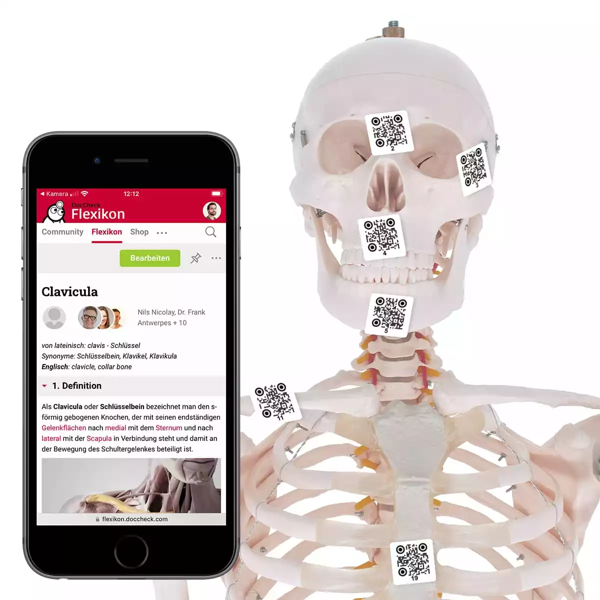
Anatomical models and teaching materials
Medical teaching materials do not only consist of texts and pictures. Turning theory into practical matters is just as important as all the theoretical learning that you do. DocCheck Shop offers you a wide range of practical teaching materials and utensils. Anatomical models of individual organs and human body sections will help you study human anatomy and training systems that give you the opportunity to practice medical procedures under realistic conditions, but completely risk-free.
Attention students and trainees! – Many offers with attractive discounts
Acquiring theoretical knowledge is never as important as during your studies. We have collected a whole range of products in the Students-Only section just for you, in order to provide you with the best help possible. These products are specially selected to suit any student’s and trainee’s needs. Did you know that you can receive up to 25% discount after registering?